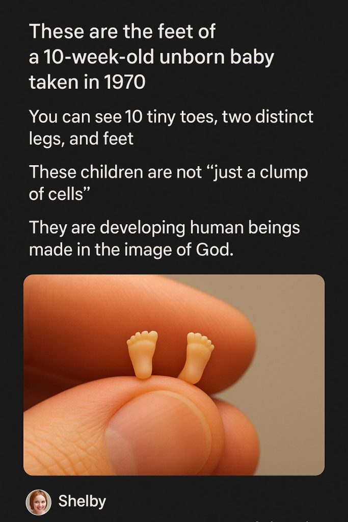
In a powerful image that has recently gone viral on social media, pro-life advocate Lila Rose shared a 1970 photograph depicting the feet of a 10-week-old unborn baby. The photo shows 10 tiny toes, two distinctive legs, and feet—serving as a striking visual reminder of fetal development taking place far earlier than some may realize.
The image challenged narratives that describe early-stage fetuses as “just a clump of cells” by presenting concrete, undeniable evidence of complex, human features already formed after less than three months of gestation.
Lila Rose, founder of the organization Live Action, has long been outspoken in highlighting scientific details about when human life begins. The recent shares and comments surrounding the image have reignited discussions about abortion, medical ethics, and the portrayal of fetal development in education and public policy.
“You can see 10 tiny toes, two distinct legs, and feet,” Lila Rose emphasized in her caption, underscoring the humanity and individuality of unborn children at this developmental milestone. This visual has been described by many online as irrefutable evidence of early life features that deserve acknowledgment in debates surrounding reproductive rights and fetal health.
Experts in embryology explain that by 10 weeks, a fetus has developed most of its vital organs and identifiable human characteristics. The hands and feet exhibit clearly differentiated toes and fingers. The external features such as eyes, ears, and limbs are becoming distinctly recognizable. This marks a critical period in prenatal development where the embryo’s transformation into a fetus is evident.
The image hails from 1970, a significant era predating the landmark 1973 Roe v. Wade Supreme Court decision that federally legalized abortion in the United States. It serves as a historical artifact showing early prenatal development long before modern ultrasound imaging further advanced public understanding.
Reactions to the photo vary broadly across the public spectrum. Supporters of pro-life movements hail the image as a compelling argument for fetal personhood and stronger protections for the unborn. On the other hand, pro-choice advocates argue for women’s rights to make reproductive choices and caution against oversimplifying complex ethical issues based solely on visuals.
Medical professionals advise that while photographs and imagery can be powerful tools for education, comprehensive understanding of fetal development and reproductive health requires nuanced discussions incorporating medical, ethical, and personal perspectives.
Even so, this 1970 photograph stands as a poignant reminder that each life’s earliest stages involve intricately formed human features, long before birth. Its viral spread reflects an ongoing societal engagement with how science and ethics intersect in conversations about life, rights, and healthcare.
As the image circulates widely through social media platforms, it continues to provoke thoughtful discourse and emotional responses. Whether viewed through a scientific lens or an ethical one, it undeniably highlights the extraordinary process of human development that takes place inside the womb.
Where to Learn More
- A Closer Look at Fetal Development at 10 Weeks – Live Action
- Fetal Development: What You Need to Know – Medical News Today
- Pregnancy and Fetal Development – Planned Parenthood
- Prenatal Development Overview – Encyclopaedia Britannica
- Ethical Considerations in Fetal Research and Rights – New England Journal of Medicine